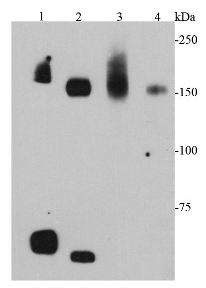
Western blot analysis of cMet on different cell lysates using anti-cMet antibody at 1/1000 dilution. Positive control: Lane 1: Mouse liver Lane 2: Mouse kidney Lane 3: D3 Lane 4: MEF ,images/20190228/48585wb-1.jpg,images/20190228/48585wb-1.jpg51545,151493,,,images/20190228/48585ihc-1.jpg,Immunohistochemical analysis of paraffin-embedded mouse liver tissue using anti-cMet antibody. Counter stained with hematoxylin.,images/20190228/48585ihc-1.jpg,images/20190228/48585ihc-1.jpg52745,151493,,,images/20190228/48585ihc-2.jpg,Immunohistochemical analysis of paraffin-embedded mouse spleen tissue using anti-cMet antibody. Counter stained with hematoxylin.,images/20190228/48585ihc-2.jpg,images/20190228/48585ihc-2.jpg53696,151493,,,images/20190228/48585ihc-3.jpg,Immunohistochemical analysis of paraffin-embedded human colon cancer tissue using anti-cMet antibody. Counter stained with hematoxylin.,images/20190228/48585ihc-3.jpg,images/20190228/48585ihc-3.jpg55193,151493,,,images/20190228/48585if-1.jpg,"ICC staining cMet in N2A cells (green). Cells were fixed in paraformaldehyde, permeabilised with 0.25% Triton X100/PBS.