免费电话:400-9961-933
邮箱:info@sabbiotech.com
欢迎光临本站
川博智能体 English
位置: 首页 > 磷酸化抗体 > MYPT1 (Phospho-Thr853) Polyclonal Antibody

MYPT1 (Phospho-Thr853) Polyclonal Antibody#12302

是否有货: 有货
产品详情

产品名称MYPT1 (Phospho-Thr853) Polyclonal Antibody

来源种属Rabbit

克隆性Polyclonal

亚型IgG

纯化The antibody was affinity-purified from rabbit antiserum by affinity-chromatography using epitope-specific immunogen.

应用WB IHC ELISA

种属反应性Human;Mouse;Rat

特异性Phospho-MYPT1 (T853) Polyclonal Antibody detects endogenous levels of MYPT1 protein only when phosphorylated at T853.

免疫原类型peptide

免疫原描述Synthesized peptide derived from human MYPT1 around the phosphorylation site of T853.

基因/蛋白名称MYPT1

标记Unconjugated

别名PPP1R12A; MBS; MYPT1; Protein phosphatase 1 regulatory subunit 12A; Myosin phosphatase-targeting subunit 1; Myosin phosphatase target subunit 1; Protein phosphatase myosin-binding subunit

数据库入口号Swiss-Prot: O14974
NCBI Gene ID: 4659

Uniprot O14974

实际分子量130kd

浓度1mg/ml

配方Liquid in PBS containing 50% glycerol, 0.5% BSA and 0.02% sodium azide.

保存Store at -20˚C/1 year

Target种属human

应用详情
Western blotting: 1/500 - 1/2000
Immunohistochemistry: 1/100 - 1/300
ELISA: 1/5000
Not yet tested in other applications
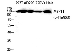
Western Blot analysis of 293T AD293 22RV1 HELA cells using Phospho-MYPT1 (T853) Polyclonal Antibody
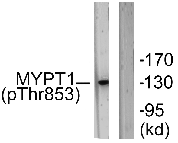
Western blot analysis of lysates from NIH/3T3 cells.The lane on the right is blocked with the phospho peptide.
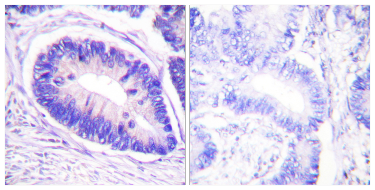
Immunohistochemistry analysis of paraffin-embedded human colon carcinoma. The picture on the right is blocked with the phospho peptide.
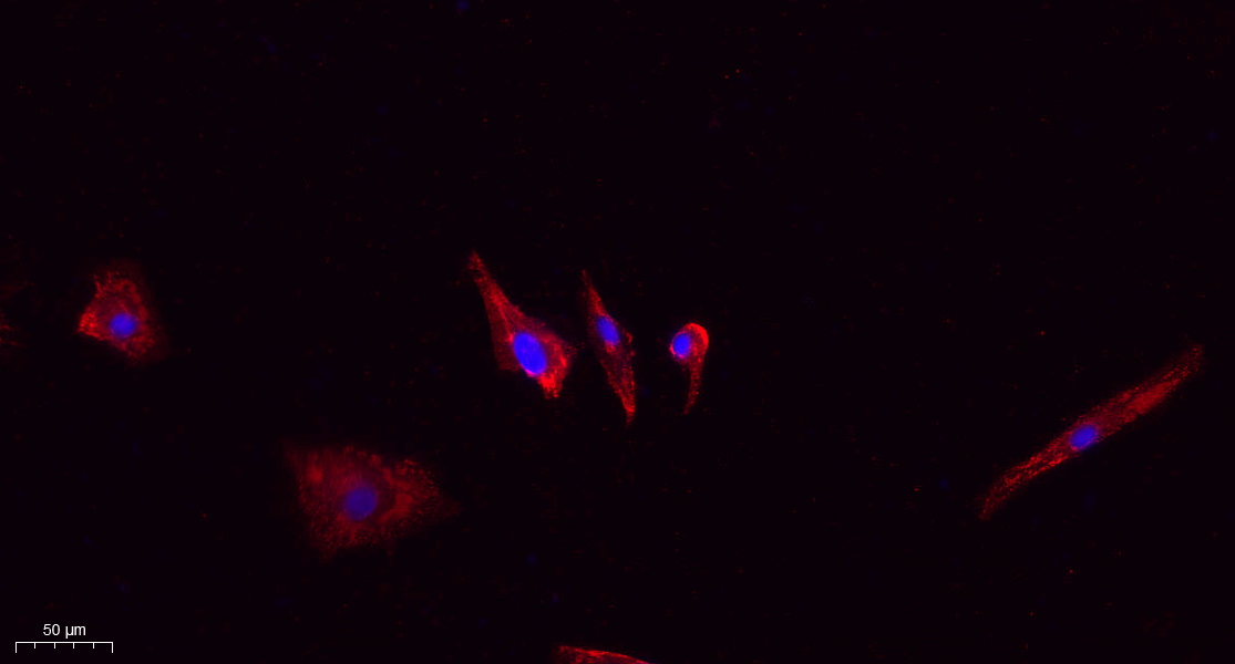
Immunofluorescence analysis of A549. 1,primary Antibody(red) was diluted at 1:200(4°C overnight). 2, Goat Anti Rabbit IgG (H&L) - Alexa Fluor 594 Secondary antibody was diluted at 1:1000(room temperature, 50min).3, DAPI(blue) 10min.
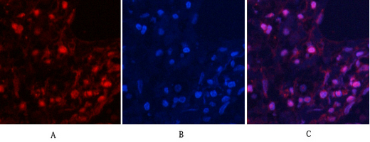
Immunofluorescence analysis of human-lung tissue. 1,MYPT1 (Phospho-Thr853) Polyclonal Antibody(red) was diluted at 1:200(4°C,overnight). 2, Cy3 labled Secondary antibody was diluted at 1:300(room temperature, 50min).3, Picture B: DAPI(blue) 10min. Picture A:Target. Picture B: DAPI. Picture C: merge of A+B
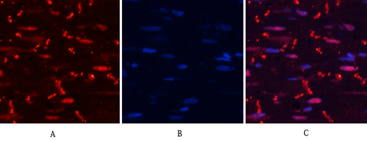
Immunofluorescence analysis of rat-heart tissue. 1,MYPT1 (phospho Thr853) Polyclonal Antibody(red) was diluted at 1:200(4°C,overnight). 2, Cy3 labled Secondary antibody was diluted at 1:300(room temperature, 50min).3, Picture B: DAPI(blue) 10min. Picture A:Target. Picture B: DAPI. Picture C: merge of A+B

如果您使用该产品12302发表了文章,请通知我们,让我们可以引用您的文献。

et al,The role of mitogen-activated protein kinase signaling pathway in bone formation during mid-palatal suture expansion , (2025),
PMID: 40298405

注释

应用

  • WB免疫印迹
  • IHC免疫组化
  • IF免疫荧光
  • ICC免疫细胞化学
  • FC流式细胞
  • IP免疫沉淀
  • E酶联免疫吸附法
  • DB免疫斑点法
  • ChIP染色质免疫沉淀
  • GICA胶体金免疫层析法
  • NC阴性对照

种属反应性

  • Hu
  • Ms小鼠
  • Rt大鼠
  • Dm果蝇
  • C线虫
  • Mk
  • Rb
  • B
  • D
  • P
  • Hm仓鼠
  • ChHm中国仓鼠
  • Chk
;

联系我们

Location :

南京市高新开发区星火路10号人才大厦C座

Phone :

400-9961-933
025-58868422

Email Address

info@sabbiotech.com